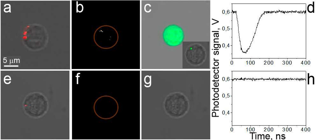
Figure 2.
Specificity of PNB mediated cargo delivery. CD3-positive (a–c) and -negative (e–g) cells in suspension. Images of target (a–c) and non-target (e–g) cells: a,e: confocal bright field and optical scattering images show NP clusters (red) in cells; b,f: time-resolved optical scattering images show bright PNB in target cell; c,g: confocal fluorescent and bright field images obtained after the PNB treatment show FITC-Dextran dye in target cell (the insert shows the initial delivery localization of dye within 10 min after PNB generation). Optical scattering time-responses show PNB-specific signal for target cell (d) and no PNB for non-target cell (h), the PNB lifetime is measured as duration of PNB-specific signals.

