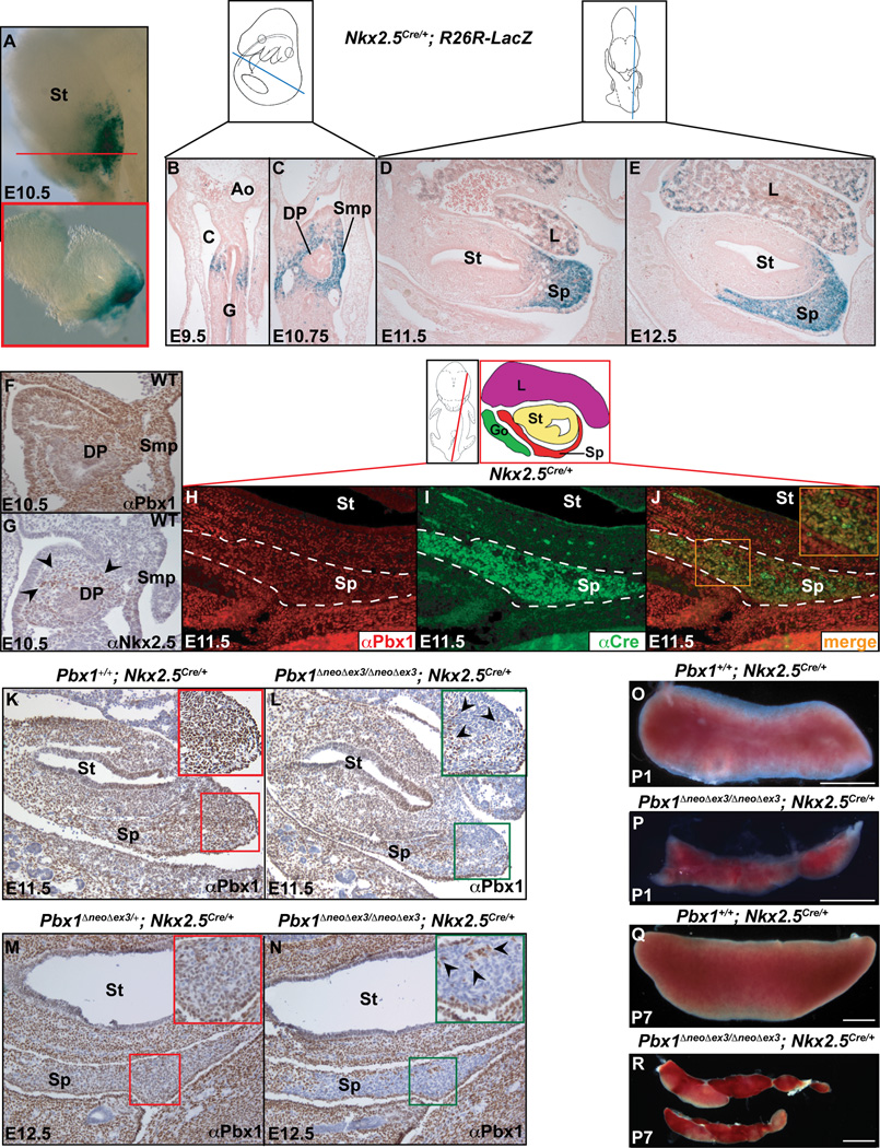
Figure 1. Pbx1 inactivation in Nkx2-5-positive mesenchyme causes spleen hypoplasia.
(A–E) Whole mount (A), transverse (A inset, red box corresponds to plane of section indicated by red line; B–C), and sagittal (D–E) sections of Nkx2-5Cre/+;R26R-LacZ embryos from E9.5 to E12.5, stained by β galactosidase. (F) IHC for Pbx1b and (G) Nkx2-5 (black arrowheads) in E10.5 WT transverse sections. (H–J) IF on E11.5 Nkx2-5Cre/+sagittal sections. Co-localization (orange, J inset) of Pbx1 (red), which is widespread in the spleen, and Cre (green) in spleen mesenchyme (outlined by white dashes). (K–N) IHC with α-Pbx1b on E11.5 and 12.5 sagittal sections from control (K,M) and Pbx1 conditional mutant (L,N) embryos, showing Cre-mediated Pbx1 loss in all but a few positive cells (black arrowheads; insets). (O–R) P1 and P7 control (O,Q) and homozygous mutant spleens (P,R). Ao, aorta; C, coelomic cavity; DP, dorsal pancreas; G, gut; Go, gonad; L, liver; Smp, splanchnic mesodermal plate; Sp, spleen; St, stomach; WT, Wildtype. See also Figures S1 and S2.

