Fig. 2.
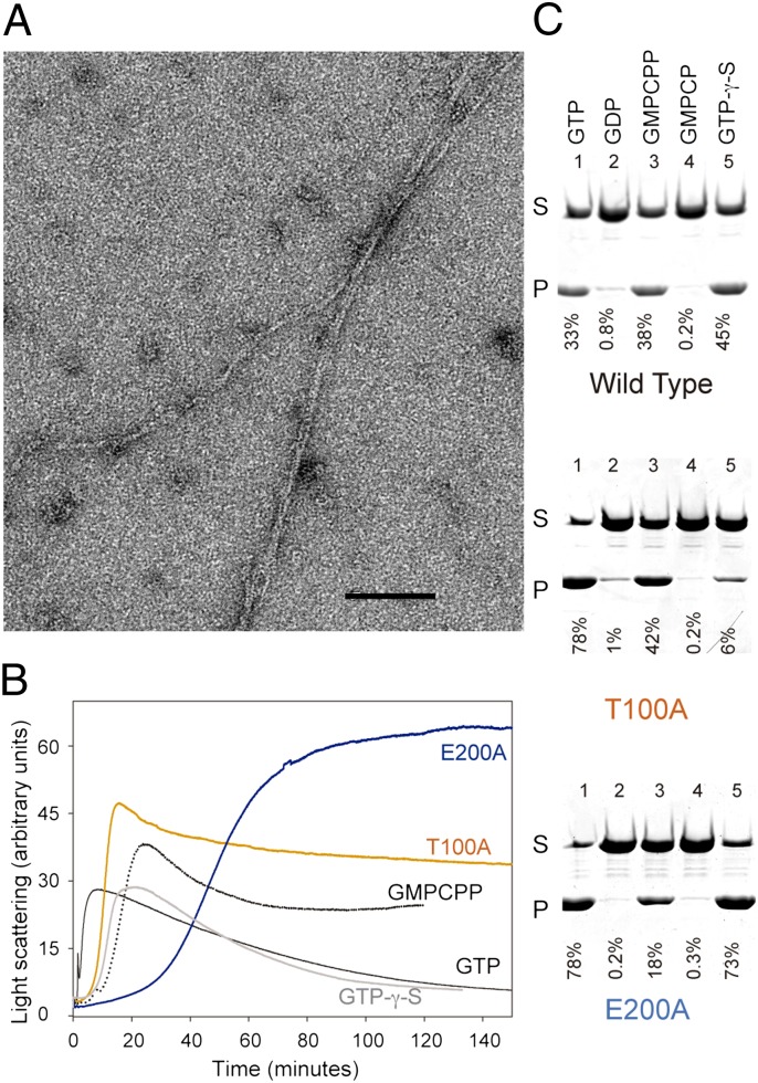
Assembly properties of phage TubZ. (A) Electron micrograph of 10 μM TubZ two-stranded helical filaments assembled in PKE buffer [50 mM piperazine-1,4-bis-2-ethanesulfonic acid–potassium hydroxide, 100 mM potassium acetate, 1 mM EDTA (pH 6.5)] with 1 mM GTP and 6 mM Mg2+. (Scale bar: 100 nm.) (B) TubZ assembly monitored by 90° light scattering. WT TubZ (10 μM) polymerized in PKE buffer with 6 mM Mg2+ and 1 mM GTP (black), 0.1 mM GMPCPP (dotted black line), or 0.1 mM GTP-γ-S (gray). TubZ-T100A (10 μM; orange) or TubZ-E200A (10 μM; blue) was assembled in PKE with 6 mM Mg2+ and 1 mM GTP. There is an increase/decrease on the signal attributable to the formation/disassembly of polymers. (C) SDS/PAGE of sedimentation assays of 10 μM TubZ (WT and mutants) assembled in PKE buffer with 6 mM Mg2+ and 1 mM GTP (line 1), 2 mM GDP (line 2), 0.1 mM GMPCPP (line 3), 0.1 mM GMPCP (line 4), and 0.1mM GTP-γ-S (line 5). P, pellet; S, supernatant. Nucleotide diphosphates GDP and GMPCP do not induce filament formation.

