Fig. 2.

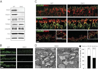
Intact cytorachitecture and synaptic morphology in retinas of mice lacking both RGS7 and RGS11. (A) Western blot analysis of protein expression in mouse retinas. Concurrent knockout of both RGS7 and RGS11 does not affect the expression of the key ON-BC signaling proteins. Retinas from four mice were used for the quantification. (B) Retinas of RGS7,RGS11 DKOs completely lack RGS7-, RGS11-, and Gβ5-positive synaptic puncta in the outer plexiform layer. (Scale bar: 10 μm.) (C) Normal morphology, dendritic branching, and accumulation of mGluR6 and TRPM1 at the dendritic tips of the ON-BC in DKO retinas. Only bipolar cells and outer plexiform layer are shown. (Scale bar: 5 μm.) (D Left) Intact morphology of synapses between rods and horizontal/bipolar cells analyzed by electron microscopy. High magnification of the DKO synapses reveals the presence of the bipolar cell (BC) and horizontal cell (HC) dendrites within the rod spherules that contained intact ribbons (*). (Scale bar: 0.5 μm.) (Right) Quantification of synapses with all three identifiable features. Quantification results are based on preparations obtained from two separate mice for each genotype. For each genotype, 200–300 synaptic triads were scored by three independent investigators for the presence of ON-BC dendrites in the rod spherules. Error bars reflect deviation of the counts among the investigators.
