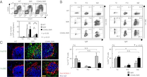
Fig. 3.

Accumulation and deletion of Sm/RNP-reactive Vκ38C+ B cells in splenic MZ in 56R mice. (A and B) Flow cytometric analysis of spleen cells from 16- to 20-wk-old WT (C57BL/6 × BALB/c) F1, 56R, and CD40L/56R mice. Cells were stained with allophycocyanin (APC)-conjugated rat anti-mouse CD21 and PE-conjugated rat anti-mouse CD23 antibody, and lymphoid gated cells were examined by flow cytometry (A). Percentages of T2, FO, and MZ B cells are indicated. Numbers of T2, FO, and MZ B cells were calculated for each spleen (Lower graph). Means ± SD of three mice are shown. Alternatively, cells were stained with Alexa Fluor 488-conjugated anti-56R/Vκ21D or anti-56R/Vκ38C anti-idiotype antibody together with APC-conjugated rat anti-mouse CD21, PE-conjugated rat anti-mouse CD23, biotinylated goat anti-mouse IgM antibodies, and PerCP-conjugated streptavidin, and lymphoid gated cells were examined by flow cytometry (B). IgM vs. 56R/Vκ21D or 56R/Vκ38C staining is displayed in T2, FO, and MZ B cells. Percentages of 56R/Vκ21D+ and 56R/Vκ38C+ cells in T2, FO, and MZ B cells were calculated. Means ± SD of three mice are shown (Lower graphs). (C) Immunohistological analysis of spleen from WT (C57BL/6 × BALB/c) F1, 56R, and CD40L/56R mice. Sections were stained for MOMA-1 (red), IgD (blue), and either 56R/Vκ21D or 56R/Vκ38C (green) and analyzed by confocal microscopy. [Scale bar (yellow line): 50 μm.] Representative data of three experiments are shown. N.S., not significant.
