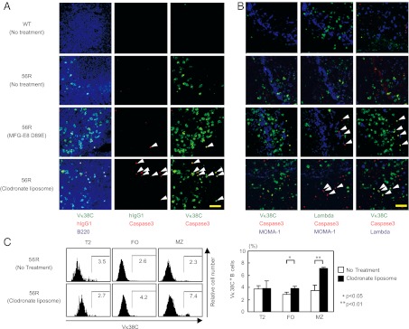
Fig. 4.

Anti-Sm/RNP Vκ38C+ B cells undergo apoptotic deletion in MZ in 56R mice. Clodronate liposome was injected i.v. to 16- to 24-wk-old 56R mice on BALB/c background at days 0, 8, 16, and 21 and mice were killed at day 23. Alternatively, MFG-E8 D89E was injected i.v. to 14- to 17-wk-old 56R mice on the BALB/c background at days 0, 3, and 6 and killed at day 7. (A and B) Spleen sections were stained for active caspase-3 (A and B), the 56R/Vκ38C idiotype (A and B), B220 (A), λ L chain (B), and MOMA-1 (B). Sections were stained with anti-human IgG1 antibody as an isotype-matched control antibody to the anti-56R/Vκ38C antibody (A). (Left, Center, and Right) The same microscopic fields viewed under different filters. Spleen sections from untreated non-Tg BALB/c mice and untreated 56R mice were examined as controls. Cells expressing both 56R/Vκ38C and active caspase-3 are indicated by arrowheads. [Scale bar (yellow line): 50 μm.] Representative data of three experiments are shown. (C) Spleen cells were stained for CD21, CD23, IgM, and 56R/Vκ38C and analyzed by flow cytometry as in Fig. 3B. 56R/Vκ38C staining is displayed in T2, FO, and MZ B cells (Left). Percentages of 56R/Vκ38C+ cells in T2, FO, and MZ B cells were calculated. Means ± SD of three mice are shown (Right).
