Fig. 2.
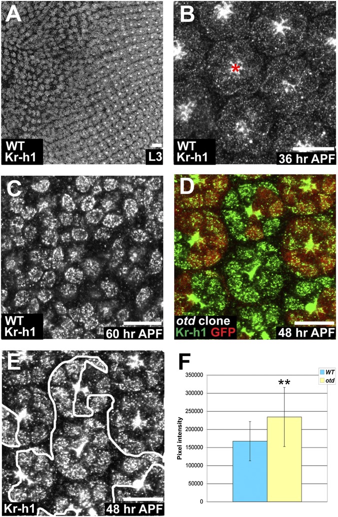
Kr-h1 expression is regulated by Otd in early pupal PRs. (A–E) (Scale bars, 10 μm.) (A–C) Kr-h1 (gray) in WT. (A) L3 eye disk. (B) 36 h APF, red asterisk (*) indicates nonspecific apical staining. (C) 60 h APF. (D) otd2 PRs. WT (red), Kr-h1 (green). (E) Kr-h1. (F) Quantification of pixel intensity of Kr-h1. WT (blue) and otd2 ommatidia (yellow). Asterisks indicate a statistically significant difference. n = 93 otd and 85 WT PR nuclei, P ≤ 10−5. Error bars represent SD.

