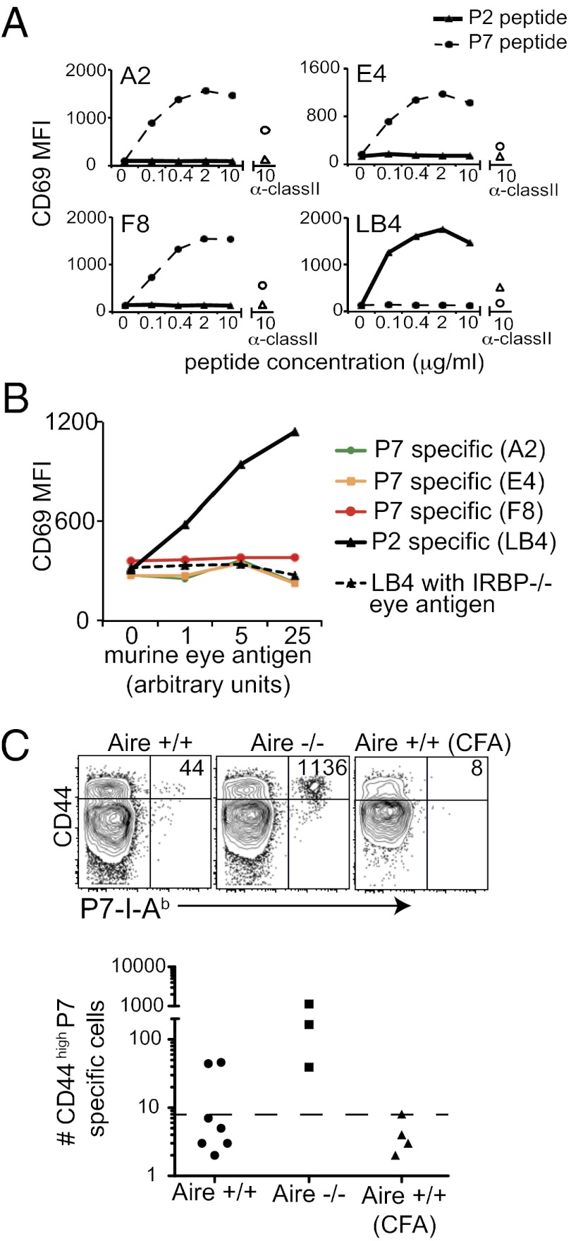
Fig. 5.
Thymic APCs preferentially present the P2 peptide epitope over the P7 peptide epitope. P7-specific (A2, E4, and F8) and P2-specific (LB4) hybridoma stimulation was measured by CD69 up-regulation. (A) T-cell hybridomas were incubated with irradiated WT B6 splenocytes and P2 or P7 peptide for 12 h. The empty triangle and circle data points indicate hybridoma stimulated in the presence of anti-class II blocking mAb plus 10 μg/mL of P2 peptide and P7 peptide, respectively. Data are representative of three independent experiments. (B) P7-specific (A2, E4, and F8) and P2-specific (LB4) hybridoma clones were incubated with CD45+CD11c+ thymic APC and soluble eye antigen from WT or IRBP−/− mice. Data are representative of two independent experiments. (C) Four- to 5-wk-old WT and Aire−/− mice were immunized with eye lysate from WT in CFA or CFA alone. At 8 d after immunization, CD44high tetramer-positive T cells in the secondary lymphoid organs were quantified. Each graphed data point represents one mouse. The dotted line in each graph is the LOD.

