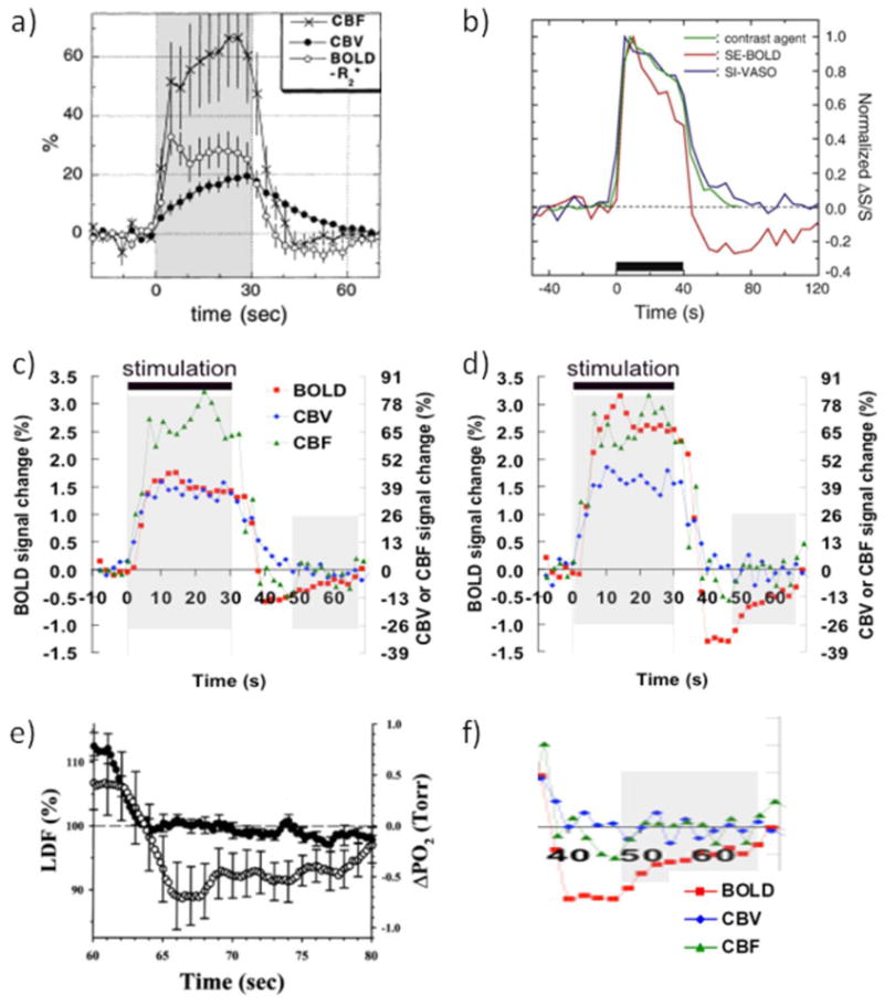
Figure 1.

(a-d) Collection of hemodynamic response data illustrating the issues surrounding the BOLD-PSU controversy. (a) GRE-BOLD, CBF and MION-CBV data from forepaw stimulation in alpha-chloralose anesthetized rats, showing delayed vascular compliance following stimulus cessation. However, also note the delayed CBV increase during stimulus onset. Reprinted by permission Reprinted from Mandeville et al., 1999a, Magn Reson Med. 42, 944, with permission from John Wiley and Sons (b) MION-CBV and VASO-CBV deep-cortex data from visual stimulation in isoflurane-anesthetized rats, showing excellent correspondence between the two methods and a rapid response to stimulus onset as well as stimulus cessation. A slower second CBV phase of much smaller magnitude than the positive response can also be seen, which returns to baseline well before the spin echo BOLD-PSU has ceased to exist. Reprinted from Jin and Kim, 2008a, Neuroimage. 40, 59 with permission from Elsevier; (c,d) Average time courses (n = 8) from human visual stimulation (yellow-blue flashing checkerboard) using VASO-CBV, CBF and GRE-BOLD for all voxels activated within a certain approach (c) and for only voxels activated in all three approaches (d), the latter reflecting better localization in parenchymal regions. Notice the lack of significant CBV and CBF delays during the large and prolonged BOLD-PSU in (d). Reprinted from Lu et al, 2004a, J. Cereb. Blood Flow Metab. 24, 764, with permission from Macmillan Publishers Ltd. (e) Laser Doppler flow and oxygen tension changes following 60 s of forepaw stimulation in halothane-anesthetized rats. Notice the lack of a flow undershoot and the prolonged undershoot in oxygen tenson of the same shape as the one found following visual stimulation in humans (d), which is show expanded in (f). Data in (e) were reprinted from Ances et al., 2008b, Neurosci Lett. 306, 106, with permission from Elsevier.
