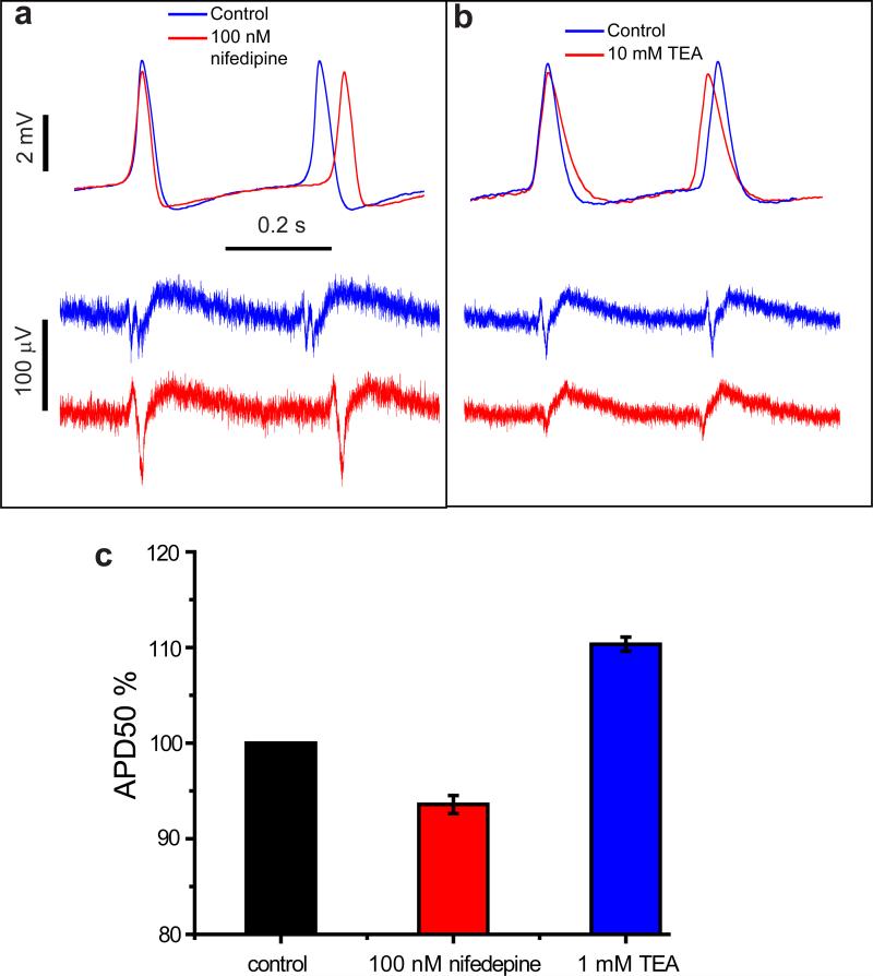
Figure 5. Effect of ion-channel blocking drugs on HL-1 cells revealed by nanopillar intracellular recording after electroporation.
With Ca2+ channel blocker nifedipine (a) and K+ channel blocker tetraethylammonium (b) administered to HL-1 cells, intracellular action potential recordings by the nanopillar electrodes (top) reveal changes in both action potential duration and period with much higher clarity than extracellular recordings (bottom). Control and drug-administered recordings are overlaid at the rising edges of the first action potential for comparison. Note the difference in vertical scale bars. (c) Statistics of nifedipine and tetraethylammonium effects on percentage change of APD50 of HL-1 cells. For each drug, 4 different HL-1 cells on 3 different cultures are measured. Note the y-axis ranges from 80% to 120% of normalized APD50. See Supplementary Table 1 and 2 for details.

