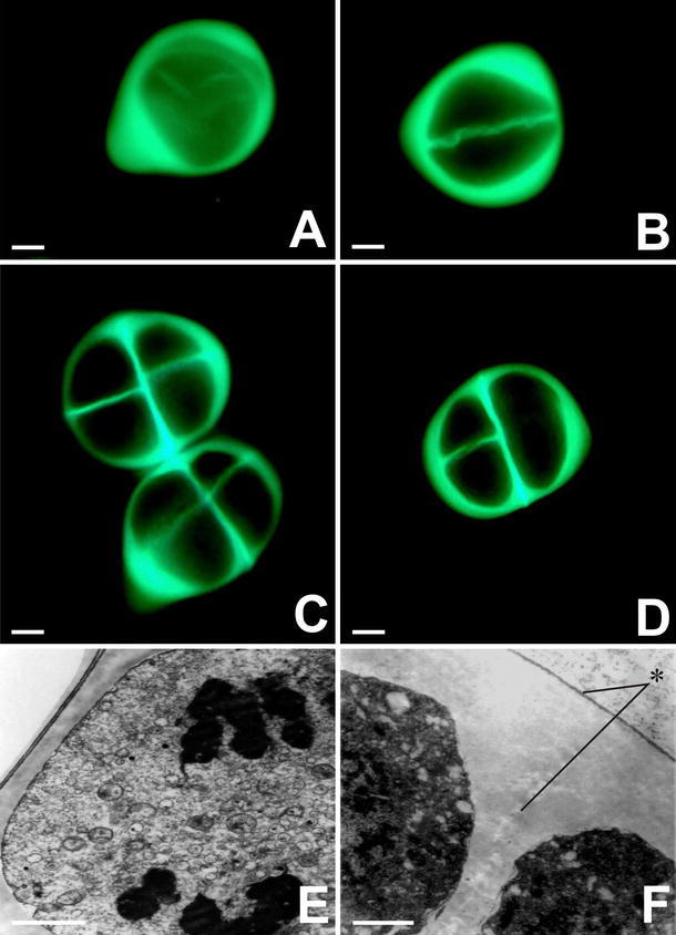
Fig. 1.

Fluorescence microscopy observation of callose deposition around a meiotic Allium sativum cell during the microsporogenesis process. a Microsporocyte. b Dyad. c Tetragonal tetrad configuration. d Decussate type. e Microsporocyte at anaphase I, surrounded by a thick callose wall. f TEM image of a fragment of the thick external wall around the microspore tetrad (asterisk). Scale bars: a–d 2 μm; e–f 10 μm
