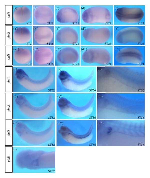
Figure 3.

Spatial expression of phd1, 2, and 3 in Xenopus embryos revealed by whole-mount in situ hybridization. (a–a′′) Lateral views with animal pole up. (b–b′′) Dorsal views with head towards left. (c–c′′) Lateral views with head towards left. (d–d′′) Ventral views with head towards left. (e–e′′) Dorsal views with head towards left. (f–g′′) Lateral views with head towards left. (h–h′′). Higher magnification views of (g), (g′), and (g′′), respectively. (i) Ventral view of (f′′) with head towards left.
