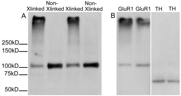
Fig. 1.
BS3 crosslinking enables the measurement of surface and intracellular pools of GluR1. A) The nucleus accumbens was dissected from a naive rat. Tissue from one hemisphere was cross-linked, whereas the other was not, generating paired samples that were then subjected to SDS-PAGE and immunoblotting for GluR1. Both high (surface-expressed) and predicted (intracellular) molecular weight bands are detected in tissue from the crosslinked side (Xlinked), whereas non-crosslinked (Non-Xlinked) tissue yields only a band corresponding to the predicted weight of GluR1. B) In crosslinked nucleus accumbens tissue, only proteins found both on the surface and inside the cell (GluR1, left) result in both high and predicted molecular weight bands. Proteins that are exclusively intracellular [tyrosine hydroxylase (TH); right] result in a predicted molecular weight band only, confirming that the crosslinker does not cross cell membranes. Reprinted from Boudreau and Wolf (2005) with permission from The Journal of Neuroscience.

