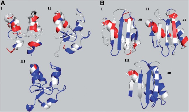
Fig. 1.

Conformations are colored according to CheShift-2. (A) Three conformations of the bovine cytochrome B5 protein are shown: 1WDB (I), rendered obsolete and replaced by 1HKO (II), both NMR-derived ensembles; and 1CYO (III) an X-ray-derived structure. (B) Three conformations of rabbit 8KDA dynein light chain protein are shown: 1BKQ (I), rendered obsolete and replaced by 1F3C (II), both NMR-derived ensembles; and 1CMI (III) an X-ray-derived structure.
