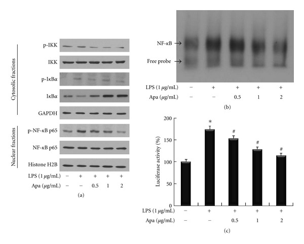
Figure 2.

The effect of apamin on NF-κB signaling pathway in LPS-treated THP-1-derived macrophages. (a) Expression levels of IKK and IκBα in the cytosolic fraction and NF-κB in the nuclear fraction were determined by western blot. GAPDH and histone H2B were used as the internal controls for cytosolic and nuclear fraction loading control, respectively. (b) Nuclear NF-κB activity was examined by EMSA. The arrow indicates the specific NF-κB band. (c) Luciferase activity was measured with a luminometer. *P < 0.05 compared to control cells, # P < 0.05 compared to control cells treated with LPS alone.
