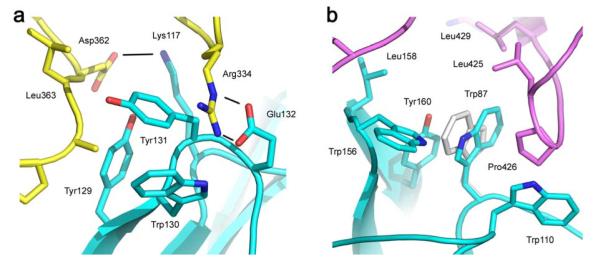
Figure 3.
Interactions at the two sub-sites. a) Two salt bridges (Arg334–Glu132; Asp362–Lys117) with three hydrogen bonds (black lines) between residues of the Cε3A domain of IgE-Fc (yellow) and the receptor (blue) contribute to sub-site 1. b) The “proline sandwich” at sub-site 2, with Pro426 in the Cε3B domain of IgE-Fc (purple) packed between Trp87 and Trp110 of the receptor (blue). The alternative orientation of Trp87 observed in the Fcε3-4/sFcεRIα complex (light grey) can be seen to make fewer contacts and a weaker interaction with Pro426.

