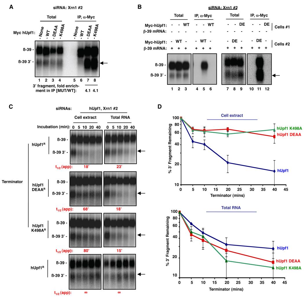
Figure 2. The 3’ NMD endonucleolytic cleavage fragment is stuck with ATPase-deficient Upf1 and is resistant to 5'-to-3' exonucleolytic decay in vitro.
(A) Northern blot for β-39 mRNA from pellet (lanes 5–8) or 5% of total extract (lanes 1–4) fractions from anti-myc IP assays from cells transiently expressing myc-tagged hUpf1 proteins indicated on the top or no exogenous protein (none). Cells were treated with Xrn1 #2 siRNA to promote the accumulation of the β-39 mRNA 3’ fragment.
(B) Same as (A), but β-39 mRNA was expressed either in the same cells as wild-type (WT) or DEAA mutant (DE) hUpf1 (lanes 2, 5, 8, 11), or in different cells and mixed prior to extract preparation (lanes 3, 6, 9, 12). Lanes 1–3, 7–9: 5% of total extracts; Lanes 4–6, 10–12: IP pellets. Lanes 1, 4, 7 and 10 are from cells not expressing Myc-hUpf1. All cells were treated with Xrn1 #2 siRNA to promote the accumulation of the β-39 mRNA 3’ fragment.
(C) Northern blots showing in vitro Terminator-mediated decay of β-39 3' mRNA fragment from extracts (left panels) or total RNA (right panels) from HeLa Tet-off cells depleted of endogenous hUpf1 using an siRNA and expressing exogenous siRNA-resistant wild-type hUpf1 (hUpf1R) or hUpf1 ATPase mutants. An siRNA targeting Xrn1 (Xrn1 #2) was included in all experiments. Time points above each lane represent the time of Terminator incubation. Bottom panels: incubation in the absence of Terminator.
(D) Quantification for each of the experiments in (C). Percent and standard deviation are calculated from three experiments.
See also Figure S2.

