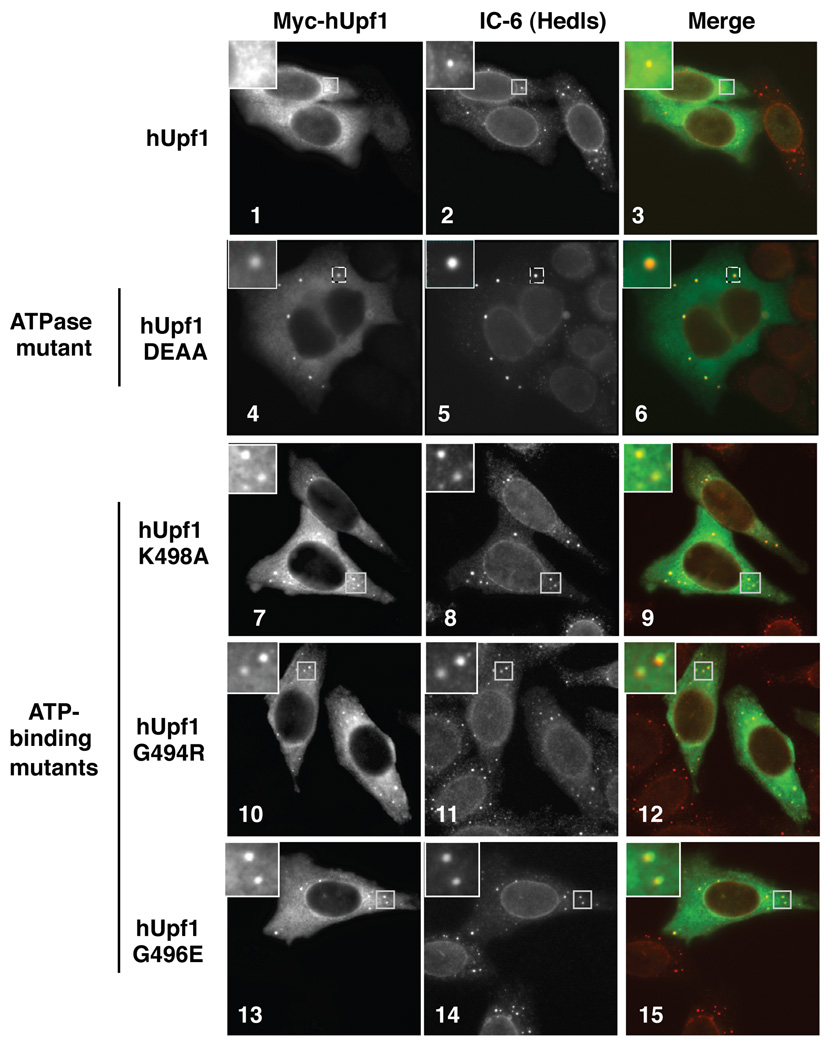
Figure 4. Mutant hUpf1 proteins deficient in ATP binding or ATP hydrolysis accumulate in PBs.
Indirect immunofluorescence assays showing localization of myc-tagged wild-type hUpf1, ATPase mutant (DEAA), or ATP-binding mutants (K498A, G494R, G496E) hUpf1 proteins transiently expressed in HeLa cells (left panels). Human IC-6 serum, which detects the decapping factor Hedls and the nuclear envelope component Lamin, was used as a PB marker (middle panels). Merged images (hUpf1: green; IC-6: red) are shown in the right panels. Enlarged images of the indicated boxed areas are shown in the upper left corner for each image.

