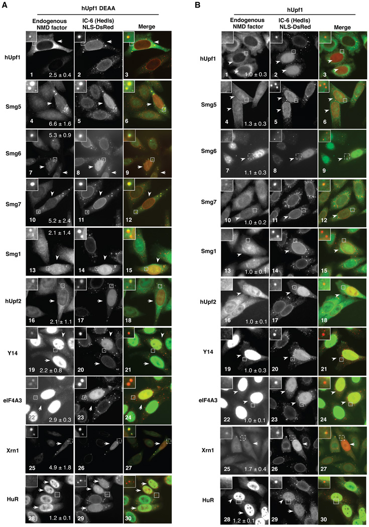
Figure 5. Multiple NMD factors accumulate in PBs in the presence of ATPase-deficient hUpf1.
(A) and (B) Indirect immunofluorescence assays showing localization in HeLa cells of endogenous NMD factors as indicated on the left, or a protein not involved in NMD, HuR, in the presence of exogenously expressed (A) hUpf1 DEAA, or (B) wild-type hUpf1. Middle panels show human IC-6 serum as a PB marker and DsRed with a nuclear localization signal to mark transfected cells (indicated by white arrowheads). Merged images (NMD factor: green; IC-6/NLS-DsRed: red) are shown in right panels. An enlarged cell section representing the boxed area of each image is shown in the upper left corner. The average enrichment of the protein factor in PBs over the general cytoplasm was quantified in transfected cells and given with standard deviation in each of the panels on the left.

