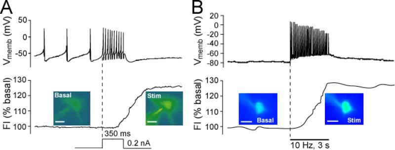
Figure 2. Basal and activity-dependent H2O2 generation in SNc DA neurons and striatal MSNs.
Representative examples of simultaneous current-clamp recordings of membrane voltage (Vmemb) and intracellular H2O2 indicated by changes in DCF fluorescence intensity (FI) in guinea-pig striatal or midbrain slices. The time course of stimulus-induced changes in DCF FI is shown with pseudocolor images recorded under basal conditions and at the end of stimulation (scale bar = 20 μm in DCF images). A) In all SNc DA neurons tested (n = 17), depolarizing current injection (0.2 nA, 350 ms) induced an increase in firing rate (upper panel) accompanied by elevated H2O2 levels (p < 0.01 vs. basal FI; lower panel). Dashed vertical line indicates onset of current injection (modified from Avshalumov and others 2005; copyright Journal of Neuroscience, used with permission). B) In all striatal MSNs tested (n = 11), local pulse-train stimulation (30 pulses, 10 Hz) generated a single action potential with each stimulus pulse (upper panel). In 7 of 11 MSNs, this activity was accompanied by a significant increase in DCF FI (p < 0.01 vs. basal) (lower panel) (modified from Avshalumov and others 2008; copyright American Physiological Society, used with permission).

