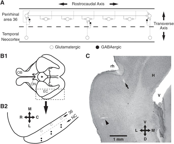
Figure 1.
Experimental setup. (A) Organization of neocortical inputs and intrinsic perirhinal connections, as inferred from previous electrophysiological and tracing studies. Dashed lines indicate neocortical axons whereas continuous lines indicate perirhinal axons. Neocortical neurons contribute stronger projections to perirhinal levels in transverse register with them where they contact both glutamatergic principal cells and feed-forward inhibitory interneurons. Neocortical neurons and principal perirhinal neurons contribute axons that course longitudinally in the perirhinal cortex. Long-range neocortical and perirhinal longitudinal axons do not contact distant local circuit cells. (B1) Ventral view of guinea pig brain. Area delimited by dashed line is expanded below. (B2) Recording sites in perirhinal area 36 (empty circles) and stimulation sites in ventral temporal neocortex (pairs of black dots). (C) Coronal section showing electrolytic lesions performed at the end of an experiment to mark a neocortical stimulation site (arrowhead) and a recording site in perirhinal area 36 (arrow). Crosses indicate orientation (R, rostral; C, caudal; M, medial; L, lateral; D, dorsal; V, ventral). Other abbreviations: EC, entorhinal cortex; H, hippocampus; NC, neocortex; OB, olfactory bulb; ON, optic nerve; rh, rhinal sulcus; V, ventricle.

