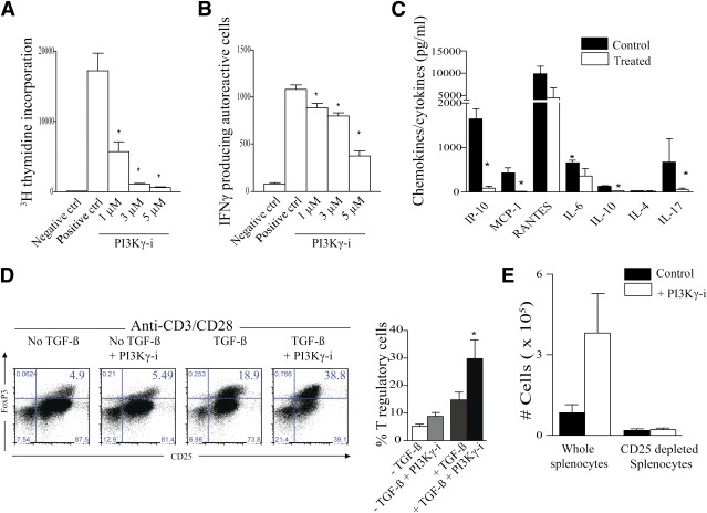
FIG. 3.
AS605240 suppressed in a dose-dependent manner the proliferation of BDC2.5 CD4+ T cells stimulated by the BDC2.5-pancreatic-peptide in vitro as measured by thymidine incorporation (A) and their production of IFN-γ in an ELISpot assay (B) (*P < 0.05, n = 3–5 mice; data are representative of three separate experiments). The negative control (ctrl) splenocytes received no peptide stimulation, whereas the positive control cells were stimulated with the BDC2.5-peptide. C: Luminex assay was used on supernatant collected from the ELISpot assay. AS605240 potently suppressed inflammatory cytokines and chemokines produced by BDC2.5 CD4+ T cells stimulated by the BDC2.5-pancreatic-peptide in vitro (*P < 0.05; n = 3–5 mice; data are representative of two separate experiments). D: Representative example of FACS staining shows the effect of the AS605240 on Treg generation in vitro using a CD3/CD28 stimulation assay with and without TGF-β. Cells are gated on CD4+ T cells. Data represent one of three separate experiments. The bar graph represents the percentage of Tregs from these experiments (*P < 0.05; n = 3–5 mice; data are representative of three separate experiments). E: Bar graph shows the absolute counts of Tregs in the spleen of NOD-scid mice that received an adoptive transfer of either whole BDC2.5 splenocytes or CD25+ depleted BDC2.5 splenocytes, followed by treatment with AS605240 for 7 days, as compared with untreated control receiving an adoptive transfer (*P < 0.05; n = 4 mice in each group). Results are presented as the mean ± SEM. (A high-quality color representation of this figure is available in the online issue.)

