FIG. 5.
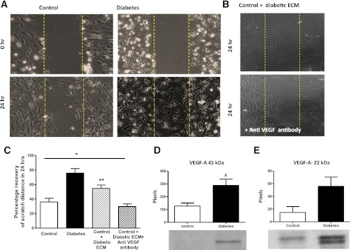
Diabetic BMECs have increased cell migration that is mediated by VEGF in an autocrine manner. A: Representative images of BMECs showing increased spontaneous cell migration. B: Representative images of control cells treated with conditioned medium from diabetic cells in the presence and absence of VEGF-neutralizing antibody. Control BMECs treated with diabetic BMEC-conditioned media show increased cell migration and anti-VEGF antibody inhibits this effect significantly. C: Quantitative analysis of data shown in A and B. Diabetic BMECs plated on fibronectin show significantly increased spontaneous cell migration after 24 h. Control cells showed enhanced migratory properties when treated with diabetic endothelial cell–conditioned media (ECM), and VEGF-neutralizing antibody significantly abrogated this response. *P = 0.0026 across control groups; **P < 0.05 vs. other control groups by Tukey post hoc analysis. Data are means ± SEM, n = 5–7. D and E: Diabetic endothelial cells secrete relatively higher levels of native and dimerized VEGF-A. #P = 0.016 diabetes vs. control. Data are means ± SEM, n = 3–8 (exact Wilcoxon test). (A high-quality color representation of this figure is available in the online issue.)

