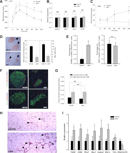
FIG. 5.
Loss of Est in ob/ob male mice aggravated diabetic phenotype, caused a loss of pancreatic β-cell mass, and increased WAT inflammation. A: GTT in ob/ob and obe male mice. B: Hepatic expression of estrogen-responsive genes in intact male mice as measured by real-time PCR analysis. C: In vivo GSIS test. D: Immunostaining of insulin and quantification of total islet area. Arrowheads indicate islets. E: β-Cell apoptosis and proliferation were measured by TUNEL assay (left) and Ki67 immunostaining (right), respectively. In both assays, the sections were counterstained with insulin. F: Immunofluorescence analysis of insulin, glucagon, and CD68 expression. G: In vitro GSIS test on isolated pancreatic islets. H: H-E staining of abdomen adipose tissue. Arrowheads indicate crownlike structures. I: Expression of macrophage markers and inflammatory genes as determined by real-time PCR. The expression of each gene was arbitrarily set as 1 in ob/ob mice. N ≥ 4 for each group. *P < 0.05; **P < 0.01; NS, statistically not significant, ob/ob versus obe. (A high-quality digital representation of this figure is available in the online issue.)

