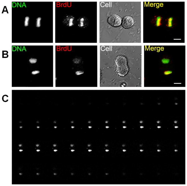
Figure 3. Asymmetric BrdU segregation at late mitotic stages.
A, A telophase cell with symmetric BrdU staining. B, An anaphase cell with asymmetric BrdU segregation. C, Z stacks of BrdU immuno-staining of B. Mitotic phases are confirmed by differential interference contrast (DIC) images, labeled as Cell.

