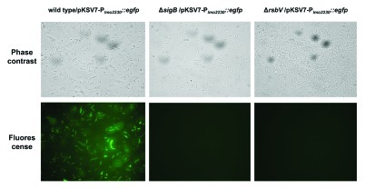
Figure 2. Fluorescence of egfp-containing cells of L. monocytogenes EGD-e wild type, ΔsigB and ΔrsbV mutants after transformation with pKSV7-Plmo2230::egfp. Phase-contrast and fluorescence microscopy of corresponding fields were performed after 1 ml of stationary phase culture was centrifuged, resuspended in 50 μl of sterile PBS and 5 μl was smeared on the slides for visualization. Images are representative of a minimum of ten randomly selected fields captured for three biological replicates for each strain.

An official website of the United States government
Here's how you know
Official websites use .gov
A
.gov website belongs to an official
government organization in the United States.
Secure .gov websites use HTTPS
A lock (
) or https:// means you've safely
connected to the .gov website. Share sensitive
information only on official, secure websites.
