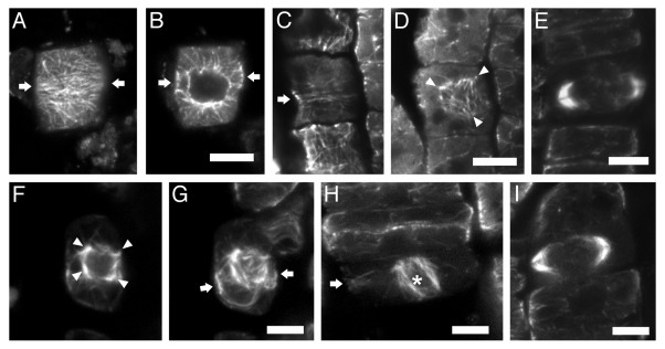
Figure 1. CLSM sections after α-tubulin immunostaining, as described in reference 3, of bot1 (A–E) and ktn1–2 (F–I) roots. The seeds of bot1 were purchased from NASC, while those of ktn1-2 mutant were offered by Dr. Masayoshi Nakamura and Prof. Takashi Hashimoto (Graduate School of Biological Sciences; Nara Institute of Science and Technology; Japan). (A, B) Early preprophase cell, at cortical (A) and intranuclear (B) section. The arrows point to the division plane. The preprophase band consists of poorly aligned microtubules (A), while the nucleus is already surrounded by numerous microtubules (B). (C and D) Late preprophase/prophase cell CLSM sections, through the cortical cytoplasm (C) and at the surface of the nucleus (D). The preprophase band is loose (C, arrow), while perinuclear microtubules exhibit various orientations (D), converging to multiple foci (arrowheads in D). (E) “Double-arrow” shaped expanding phragmoplast in cytokinetic bot1 cell. (F and G) Late preprophase/prophase cell at intranuclear CLSM section (F) and maximum projection of 8 successive sections (G). The arrows in (G) point to the division plane. The perinuclear microtubules exhibit various orientations, converging to multiple foci (arrowheads in F). (H) Prometaphase cell (the asterisk indicates the spindle) with preprophase band remnants at the cell cortex (arrow). (I) “Double-arrow” shaped expanding phragmoplast in cytokinetic ktn1–2 cell. Scale bars: 5 μm.

An official website of the United States government
Here's how you know
Official websites use .gov
A
.gov website belongs to an official
government organization in the United States.
Secure .gov websites use HTTPS
A lock (
) or https:// means you've safely
connected to the .gov website. Share sensitive
information only on official, secure websites.
