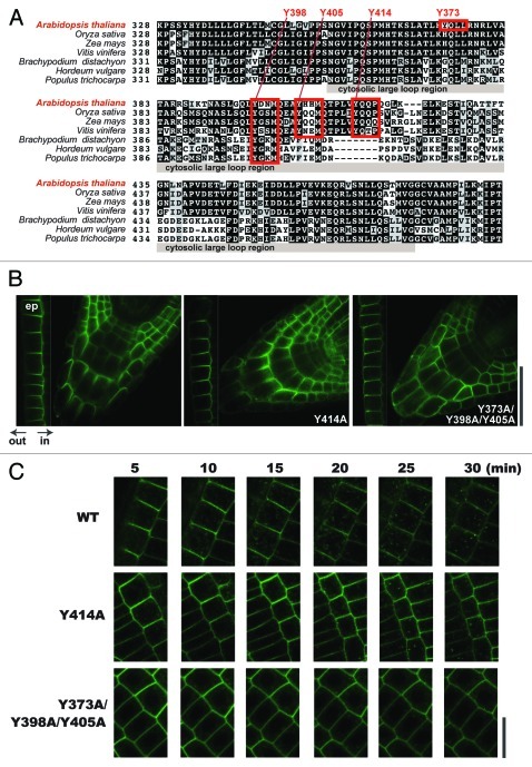
Figure 1. Effect of Y414A substitution on the polar localization and endocytic degradation of BOR1. (A) Amino acid sequences of the cytosolic large loop region of BOR1 and its homologs. The sequences were aligned using the Clustal W program (clustalw.ddbj.nig.ac.jp/). Putative tyrosine-based signals are highlighted. (B) Involvement of tyrosine-based signals in polar localization of BOR1. BOR1GFP with tyrosine–to–alanine substitution in elongating epidermal cells (ep; left end of each panel) and root tips under low B conditions. Transgenic lines expressing BOR1GFP(WT), BOR1(Y414A)GFP, and BOR1(Y373A/Y398A/Y405A)GFP under control of the BOR1 promoter are shown. Plants were grown on vertically placed solid medium containing 0.3 µM boric acid, 1% sucrose, and 1.5% Gellan gum (Wako Pure Chemicals) for 5 d. Other growth conditions were the same as in the previous work.4 (C) Time-course analysis of the subcellular localization of BOR1GFP(WT), BOR1(Y414A)GFP, and BOR1(Y373A/Y398A/Y405A)GFP after high B supply. Plants were grown on solid medium containing 0.3 µM boric acid for 5 d. The plants were then transferred to solid medium containing 100 µM boric acid and 0.5% Gellan gum. Time after shifting to high B medium is indicated. Laser scanning confocal microscopy was performed using a Zeiss LSM510 META. Excitation and detection wavelengths for GFP were 488 nm and 505–530 nm (band path), respectively. Scale bars indicate 50 µm (B) and 20 µm (C), respectively.

An official website of the United States government
Here's how you know
Official websites use .gov
A
.gov website belongs to an official
government organization in the United States.
Secure .gov websites use HTTPS
A lock (
) or https:// means you've safely
connected to the .gov website. Share sensitive
information only on official, secure websites.
