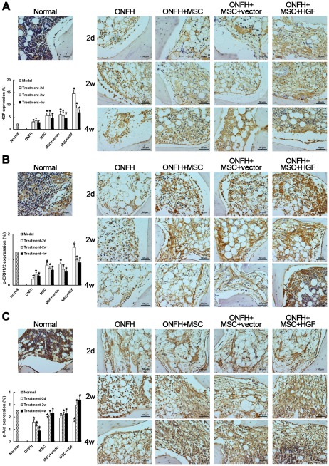
Figure 7. Immunohistochemical detection and semi-quantitative analysis of HGF expression (A), phosphorylation of ERK1/2 (p-ERK1/2) (B) and Akt (p-Akt) (C).
There was a little increase in HGF after trauma. After the transplantation of MSCs, the HGF level increased significantly at as early as 2 days, which was concomitant with increased p-ERK1/2. The HGF level decreased gradually for 2 weeks after transplantation, followed by a significant increase in Akt activation. The effects were most marked in the animals treated with HGF-transgenic MSCs. * P<0.05, compared with the normal group. # P<0.05, compared with the non-infected MSC-treated group. Scale bar = 50 µm.

