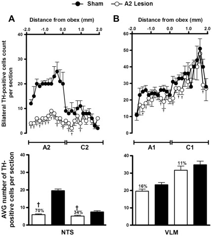
Figure 2. Lesion of A2 noradrenergic neurons with nanoinjections of anti-DβH-saporin into the NTS.
Number and average (mean ± S.E.M.) of TH-positive cells in 40-µm-thick sections from the dorsal (A) and the ventrolateral medulla (B). Sections were taken from 1.9 mm rostral to the obex to 1.9 mm caudal to the obex in animals submitted to A2 or sham lesions. Bilateral nanoinjections of anti-DβH-saporin into the NTS produced a loss of TH-containing neurons in this area (A2 group, loss = 70%; C2 group, loss = 34%), in the RVLM (C1 group, loss = 11%) and in the CVLM (A1 group, loss = 16%). † p<0.05 compared with sham lesion.

