
Figure 3.
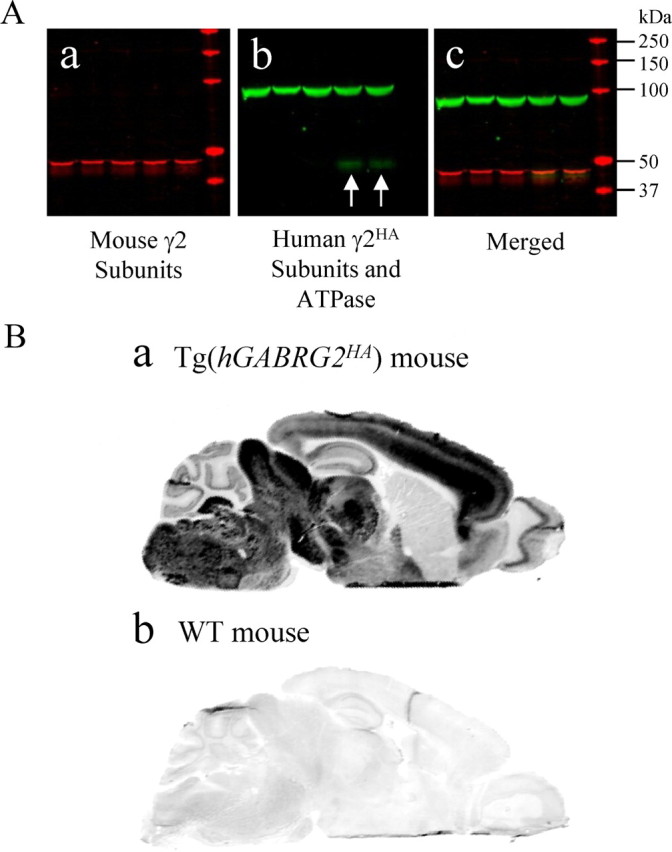
The wild-type human hGABRG2HA BAC in transgenic mouse brain had the same expression pattern as the endogenous mouse mGABRG2. A, Western blot on brain total lysate of Tg(hGABRG2HA) transgenic mice showing transgenic mice expressed both endogenous mouse γ2 subunits and HA-tagged human γ2 subunits (n = 4). a, Endogenous mouse γ2 subunits were labeled in the red channel. b, ATPase and HA-tagged proteins were labeled in the green channel. c, The merged image showed the molecular size of HA-tagged human γ2 subunits and endogenous mouse γ2 subunits were similar. The white arrows in b point to the HA bands. B, The expression pattern of γ2HA subunits in the Tg(hGABRG2HA) mouse brain was similar to that of γ2 subunits in wt mouse brain. HA antibodies stained parasagittal sections of adult Tg(hGABRG2HA) BAC transgenic mouse brain (a) or adult wild-type littermate brain (b). Sections were scanned in Odyssey scanner as one image after immunolabeling, which was presented in grayscale. The signal in the Tg(hGABRG2HA) section was oversaturated in some regions because the setting was chosen to visualize the nonspecific binding in the wild-type littermate section (n = 3).