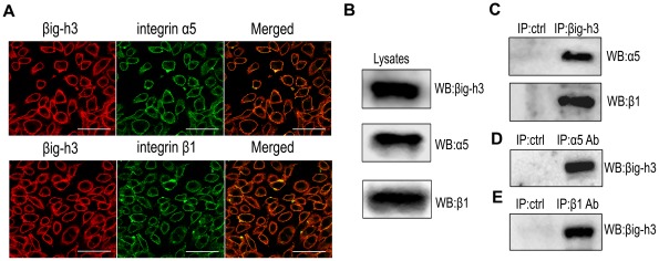
Figure 3. Expression and immunoprecipitation of βig-h3 and integrin α5β1 in U87 cells.
(A) U87 cells were double-stained for βig-h3 (red) and integrin α5 or β1 (green). (B) Expression of βig-h3 and integrin α5 and β1 subunits in U87 cell lysates. (C) Precipitates from βig-h3 immunocomplexes were detected for precipitated integrin α5 and β1 subunits. Mouse IgG was used as a negative control. Precipitates from α5 (D) or β1 (E) immunocomplexes were detected for precipitated βig-h3. Mouse IgG was used as a negative control. Bar = 50 um.

