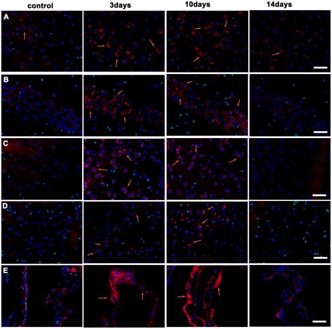
Figure 5. Detection of ATP7A by immunofluorescent staining (red) in the coronal brain slices of tx mice.
Tx mice were subjected to PA administration for 3 days, 10days and 14days, respectively. (A) Internal pyramidal layer. ATP7A immunoreactivity staining was weak in internal pyramidal layer neurons in the control mice cortex, but increased significantly on the 3rd and the 10th day, then decreased on the 14th day. (B)hippocampus CA1 region, (C)caudate putamen(CPu), (D) central medial thalamic nucleus(CM),(E)choroid plexus in the lateral cerebral ventricles.ATP7A staining was weak in cells of hippocampus CA1 region, CPu, CM and choroid plexus in the control mice, and significantly increased on the 3rd day and the 10th day during PA administration, and declined on the 14th day. Scale bars = 30 um.

