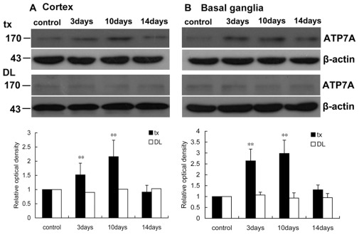
Figure 10. Western blot analysis of ATP7A in the cortex and basal ganglia of tx and DL mice.
(A) ATP7A expression increased significantly (P<0.01) on the 3rd day and 10th day in tx mouse cortex during PA treatment, and declined closed to the level of the control on the 14th day. ATP7A expression did not change in DL mice during PA treatment. (B) ATP7A expression increased significantly (P<0.01) on the 3rd day and 10th day in tx mouse cortex, and declined closed to the level of the control on the 14th day, while it did not change in DL mice. Relative protein quantified as compared with tx or DL controls (normalized to 1.0). **p<0.01 was considered significantly different from the control.

