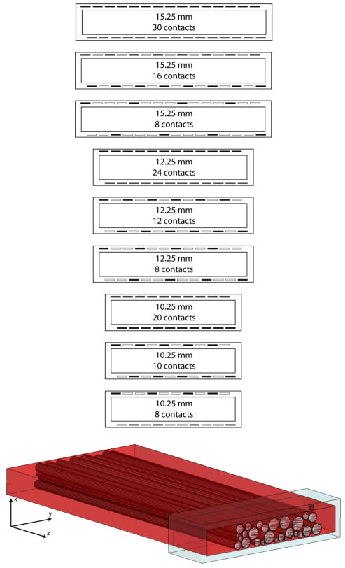
Fig. 1.
(top) The distribution of contacts within the different modeled FINEs. The opening height of the cuff was varied from 2–3 mm. The opening width of the cuff was varied from 10.25–15.25 mm. The number of contacts in the cuff was varied from 8–30. Dark rectangles in the cuff indicate the location of contacts. (bottom) The 3D FEM model derived from a human sciatic nerve histological cross section. The model contains a plane of symmetry so all objects were split across the z-axis. This included splitting the cuff, the contact seen in the upper right corner of the cuff, all neural tissue, and the saline environment (not shown).

