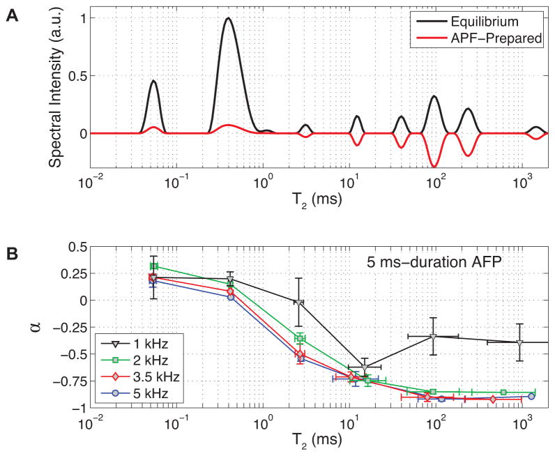
FIGURE 4. Observed effects of a sech AFP pulse on cortical bone water longitudinal magnetization.
T2 spectra (A) from a representative bone specimen at equilibrium and following an AFP pulse (5ms/5kHz) show a largely saturated bound water component (T2 ≈ 0.4 ms) and inverted pore water (T2 > 1 ms), as represented by the negative spectral amplitudes (see Methods for details). The ratio of AFP-prepared to equilibrium T2 spectral peak areas gives the APF efficiency parameter α, shown in B for a variety of AFP bandwidths (error bars represent ±1 SD across specimens).

