
Figure 1.
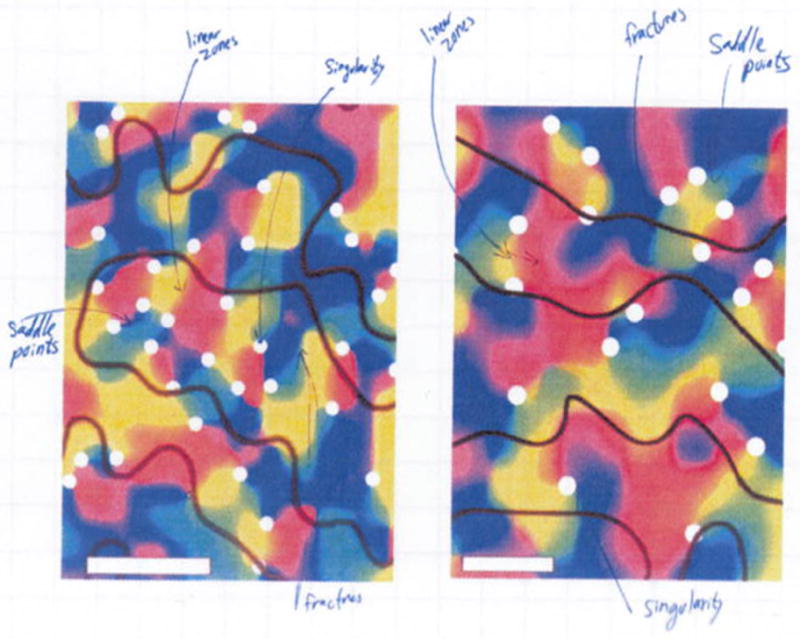
is a snapshot from Noam Harel’s notebook (early 2006) showing two of the initial orientation fMRI maps obtained in humans. The maps exhibits the spatial features that have been reported in animal studies, such as linear zone, singularities, saddle points and fractures following (Obermayer and Blasdel, 1993).