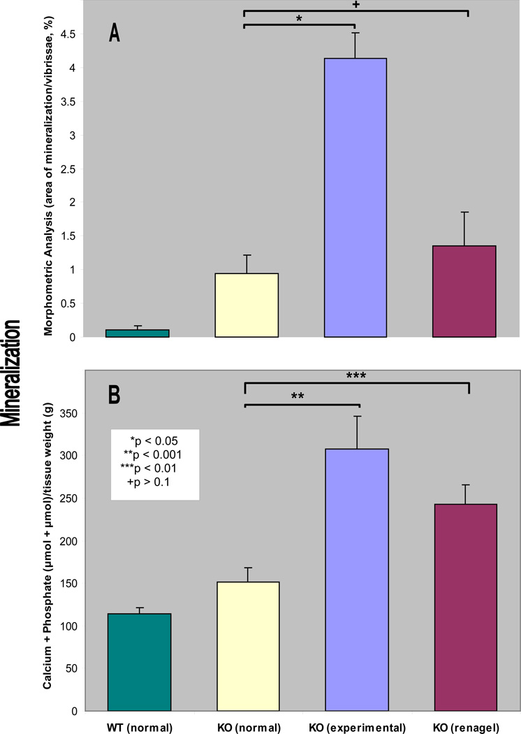
Figure 2.
Quantitation of the mineralization either by using computerized morphometric analysis, as shown in Fig. 1 (A), or by chemical determination of calcium plus phosphate content in the muzzle tissue containing the vibrissae (B). The bars represent mean ± S.E., n = 4–5 in each group. Note the statistical significance, as indicated in an inset in B.

