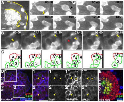
Fig. 1.
Primordial germ cell migration occurs during endodermal epithelial remodeling and ingression. Drosophila embryos imaged dorsally with anterior to the left. (A,B) Still images from a time series (supplementary material Movies 1-3). Embryos are derived from Pnos::egfp-moe::nos 3′UTR-expressing mothers and carry zygotic copies of this transgene. (A) Endodermal epithelium is outlined in yellow. Arrow highlights a migrating primordial germ cell (PGC). Still images demonstrate that PGCs do not deform as they migrate. (B) During endodermal remodeling, cells acquire a rounded-up phenotype and delaminate from the epithelium (red arrowhead). Time is shown in minutes:seconds (C) Illustration of cell identity and position in time series in B. PGCs (1-4) are outlined in green. Black arrowheads highlight PGC 3 (same as yellow arrowhead in B), which migrates between endodermal cells A and B (red). (D) Mid-stage 10 insc-lacZ embryo. The boxed regions 1 and 2 are magnified on the right. β-gal-positive cells (green in D, grayscale in 1′ and 2′) are ingressing and appear rounded or spindle shaped (yellow arrowhead in 2; phalloidin is blue in D, grayscale in 1″ and 2″). aPKC antibody staining appears discontinuous (aPKC is red in D, grayscale in 1″′ and 2″′). A migrating PGC is identifiable by enriched phalloidin staining (yellow asterisk). (E) By late stage 10, cells expressing β-gal (green) have ingressed (endoderm marked with DHB9 antibody, red). PGCs have migrated out of the endoderm (Vasa antibody in blue). Scale bars: 10 μm in A,B; 20 μm in D.

