Fig. 2.
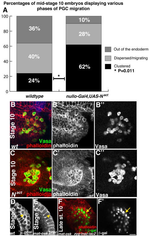
Delaying epithelial remodeling delays PGC migration. Drosophila embryos imaged dorsally (B,C), posteriorly (D,E) or laterally (F) with anterior to the left. (A) Analysis of PGC migration at stage 10 in wild-type embryos versus nullo-Gal4,UAS-Nact embryos, in which interstitial cell precursors (ICPs) are not specified. Twenty-four percent (6/25) of wild-type embryos show PGCs inside the endoderm as compared with 62% (18/29) of nullo-Gal4,UAS-Nact embryos, a statistically significant difference (*P=0.011, two-tailed Fisher’s exact test). (B-B″) A stage 10 wild-type (wt) control embryo. PGCs labeled with an antibody to Vasa (green in B, grayscale in B″) have initiated migration and the endoderm is no longer epithelial (phalloidin is red in B, grayscale in B′). (C-C″) A stage 10 nullo-Gal4,UAS-Nact embryo. PGCs (labeled with Vasa antibody, green in C, grayscale in C″) have not initiated migration and the endoderm retains epithelial character (phalloidin is red in C, grayscale in C′). (D) PGCs (arrowhead) at the posterior pole of a stage 5 embryo stained with phalloidin. (E) A stage 5 embryo from an osk54/osk301 mother lacks PGCs (arrowhead; stained with phalloidin). (F,F′) A late stage 10 embryo laid by an osk54/osk301 mother. In the absence of PGCs, insc-lacZ cells ingress (arrow; β-gal antibody, green in F, grayscale in F′; phalloidin in red). Scale bars: 20 μm.

