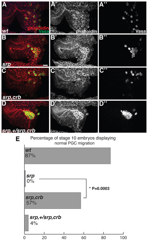
Fig. 3.
Epithelial disruption in the absence of endodermal specification is sufficient for PGC migration. (A-D″) Drosophila embryos imaged laterally with anterior to the left. Late stage 10 embryos stained with phalloidin (red) and Vasa antibody (green). (A-A″) In wild-type embryos the endoderm has lost epithelial character and PGCs have initiated migration. (B-B″) In srp mutants PGCs are unable to migrate and are surrounded by epithelial tissue. (C-C″) In srp,crb double mutants PGCs appear to migrate and are surrounded by tissue that has lost epithelial character. (D-D″) Embryos carrying the original srp mutation over the srp,crb recombinant chromosome phenocopy srp mutants (C) in both the epithelial character of the tissue surrounding PGCs and the inability of PGCs to migrate. (E) The percentage of embryos displaying normal migration, as characterized by the absence of clusters of eight or more PGCs, was 87% (55/63) for wild type, 0% (0/16) for srp and 57% (13/23) for the srp,crb double mutant. The difference between srp and srp,crb was significant (*P=0.0003, two-tailed Fisher’s exact test). The percentage for the original srp mutation over the srp,crb recombinant chromosome was 4% (1/23). Scale bar: 20 μm.

