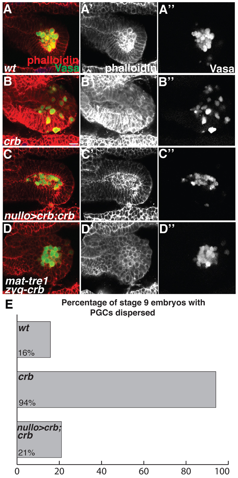
Fig. 4.

Premature epithelial disruption leads to precocious PGC migration. (A-D″) Stage 9 Drosophila embryos imaged laterally with anterior to the left and stained with phalloidin (red) and Vasa antibody (green). (A-A″) In wild-type embryos the tissue surrounding PGCs is epithelial and PGCs are clustered. (B-B″) In crb mutant embryos the tissue surrounding PGCs is not epithelial and PGCs appear to be migrating. (C-C″) Expressing UAS-crb in somatic tissue with nullo-Gal4 in a crb mutant background restores the epithelial character of the tissue and blocks premature migration. (D-D″) In crb mutant embryos derived from tre1 mutant females the tissue is not epithelial; however, PGCs do not migrate out of the endoderm. (E) The percentage of stage 9 embryos with dispersed PGCs (genotypes as in A-C). In 16% (3/19) of wild-type embryos PGCs were dispersed, whereas 94% (16/17) of crb embryos had PGCs dispersed. In nullo-Gal4,UAS-Crb,crb embryos, 21% (4/19) had PGCs dispersed. Scale bar: 20 μm.
