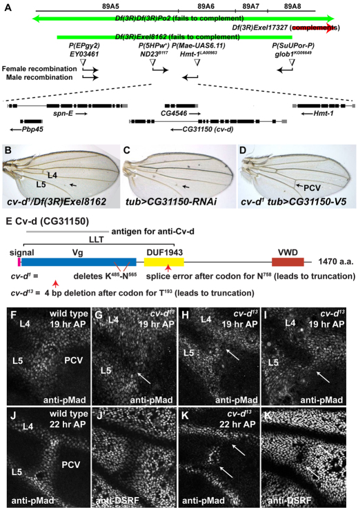
Fig. 1.
cv-d mutants disrupt CG31150 and reduce BMP signaling in the developing PCV. (A) Mapping of cv-d1 to five candidate genes, using molecularly defined Exel deletions (Parks et al., 2004), recombination relative to w+ P element insertions in w; cv-d1 Pr/P element females and transposase-mediated recombination at P element insertions (Chen et al., 1998) in Δ2-3; Gl cv-d1 Pr/P element males. (B) Disruption of PCV (arrow) in cv-d1/Df(3R)Exel8162 adult wing, normally found between longitudinal veins 4 and 5 (L4 and L5). (C) Disruption of PCV (arrow) in tub-gal4 UAS-CG31150-RNAi (VDRC #3975) adult wing. (D) Rescue of PCV (arrow) in cv-d1 homozygote by tub-gal4 UAS-CG31150-V5.(E) Structure of Cv-d, and predicted alterations in cv-d1 and cv-d13 mutants (supplementary material Fig. S1A). (F-K’) Comparison of BMP signaling in PCV regions of 19 hour AP (F-I) and 22 hour AP (J-K’) wild type (F,J,J’) and cv-d13 (G-I,K,K’) wings. Anti-pMad staining is reduced and anti-DSRF staining heightened in regions of the cv-d13 PCV (arrows).

