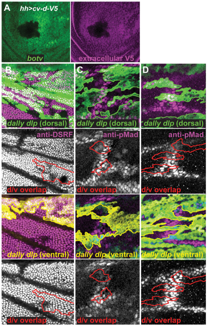
Fig. 4.
HSPGs interact with Cv-d and affect PCV development. (A) Extracellular Cv-d-V5 (purple) in wing disc is reduced in botv clone (absence of green GFP). (B-D) Dorsal and ventral epithelia around pupal PCV with clones lacking dally and dlp (absence of green or yellow GFP). Green and yellow outlines show dorsal and ventral clones, respectively; red outlines show overlapping regions missing dally and dlp on both surfaces (d/v overlap). PCV, which is marked by the suppression of DSRF (G; purple and white) or the presence of pMad (H,I; purple and white), forms in cells two to three cell diameters distant from wild-type cells in the same or the opposite epithelium.

