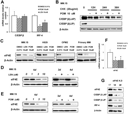
Figure 4.
IMiD compounds down-regulate C/EBPβ by targeting its protein translation. (A) MM.1S cells were cultured with pomalidomide, lenalidomide, or 0.01% DMSO (control). Total RNA was extracted from 12-hour cultures, reverse transcribed to cDNA, and used for quantitative real-time PCR. Data were analyzed according to the ΔΔCt method. Results are shown as mRNA expression relative to control (DMSO). mRNA levels were normalized with β-actin mRNA expression as control. (B) MM.1S cells were incubated with pomalidomide (10μM) with and without cycloheximide (20 μg/mL) for 12, 24, or 36 hours. Cells were lysed, and whole cell lysates were analyzed by Western blotting for C/EBPβ. (C) MM.1S, H929, OPM2, or primary myeloma cells were cultured with lenalidomide, pomalidomide, or 0.01% DMSO as control for 3 days. Cells were lysed, and whole cell lysates were analyzed by Western blotting for eIF4E. β-Actin expression was probed for loading control. (D-E) MM.1S cells (2 × 10⁶) were incubated with lenalidomide (D) or pomalidomide (E) at different concentrations with a fixed time period of 4 days, or for different time periods with fixed concentration of 10μM, or with DMSO 0.01% as control. Cells were lysed, and whole cell lysates were analyzed for eIF4E expression by Western blotting. β-Actin expression was probed for loading control. (F) MM.1S cells were incubated with lenalidomide or pomalidomide for 12 hours, and total RNA was extracted by Trizol and followed by real-time PCR. Data were analyzed according to the ΔΔCt method. Results are shown as mRNA fold compared with control (DMSO). (G) eIF4E knockdown cell lines were generated by lentiviral infection of MM.1S cells. 1# and 2# indicate different eIF4E shRNA sequences. Green fluorescence protein was used as control for eIF4E shRN-expressing cells. Cell lysates were analyzed by Western blotting to compare the levels of eIF4E, C/EBPβ, and IRF4. β-actin expression was probed for loading control.

