
Figure 1.
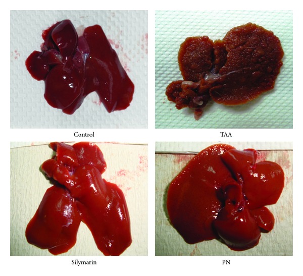
Images showing the macroscopic appearances of livers from different experimental groups. (Control): regular smooth surface. (Hepatotoxic): iirregular whitish micro- and macronodules and a large area of ductular cholangiocellular proliferation embedded within fibrosis. (Silymarin): smooth surface. (High dose PN): nearly smooth surface.