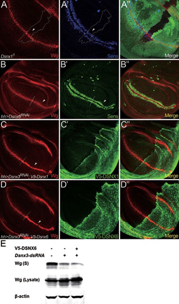
Figure 6.
DSNX1 and DSNX6 are not required for Wg secretion and signaling. (A-C) Wing discs are oriented dorsal top-right, anterior top-left. (A-A'') Wg and Sens staining in a wing disc bearing Dsnx11 clones. Mutant clone is outlined by the dotted lines to mark the absence of GFP (A'', green). Wg levels were not altered in the absence of Dsnx1 (arrowhead in A). Sens staining was also not altered in the mutant clone (arrowhead in A'). (B-B'') UAS-Dsnx6RNAi was expressed in wing discs using hhGal4 to deplete Dsnx6 activity in the P compartment. A/P boundary is shown by the dotted line. Wg was secreted normally under these conditions (arrowhead in B), and the expression of Sens was also not reduced (arrowhead in B'). (C-C'') UAS-Dsnx3RNAi and UAS-V5-Dsnx1 were co-expressed in the P compartment using hhGal4. Wg accumulated in the P compartment (arrowhead in C), indicating that the Wg secretion defect caused by DSNX3 depletion cannot be rescued by the overexpression of DSNX1. (D-D'') UAS-Dsnx3RNAi and UAS-V5-Dsnx6 were co-expressed in the P compartment using hhGal4. Wg accumulated in the P compartment (arrowhead in D), indicating that the Wg secretion defect caused by DSNX3 depletion cannot be rescued by the overexpression of DSNX6. (E) The amount of Wg in the supernatant (S) was strongly reduced when S2+pMK-Wg cells were treated with dsRNAs targeted to the coding region of Dsnx3. However, the Wg secretion defect caused by DSNX3 depletion cannot be rescued by the overexpression of DSNX6.

