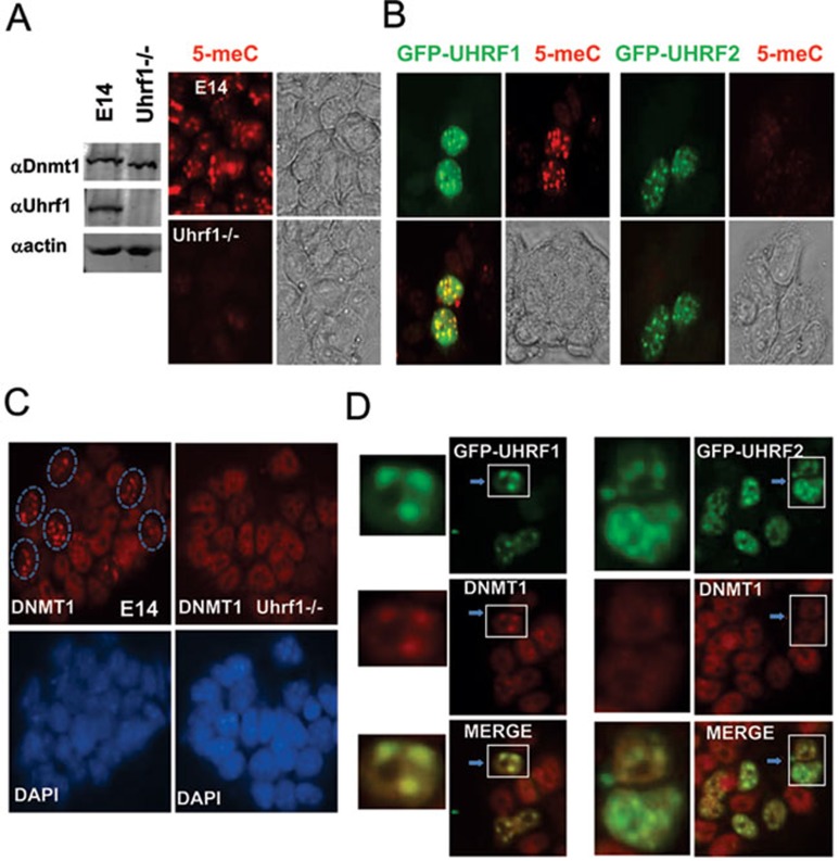
Figure 7.
Overexpression of UHRF2 in mouse Uhrf1−/− ES cells rescues neither DNA methylation defect nor pericentric heterochromatin localization of DNMT1 in S phase. (A) Confirmation of DNA methylation defect in Uhrf1−/− ES cells by anti-mC immunostaining. Left panel shows western blot analysis of whole cell extracts derived from wild-type E14 and Uhrf1−/− ES cells. Right panel shows representative 5-meC immunostaining data for E14 and Uhrf1−/− ES cells. Also shown are the phase contrast images. (B) Ectopic expression of GFP-UHRF1 but not GFP-UHRF2 rescued DNA methylation defect in Uhrf1−/− ES cells. Note both GFP-UHRF1 and GFP-UHRF2 exhibited a focal staining pattern, in agreement with their expected pericentric heterochromatin localization. (C) Immunostaining for DNMT1 confirmed absence of focal staining pattern (pericentric heterochromatin targeting) for DNMT1 in Uhrf1−/− cells. The cells with DNMT1 focal staining in E14 were circled. Also shown are DAPI staining images. (D) Expression of GFP-UHRF1 but not GFP-UHRF2 restored correct pericentric heterochromatin targeting of DNMT1. The Uhrf1−/− ES cells were transfected with GFP-UHRF1 or GFP-UHRF2 and then subjected to immunostaining for DNMT1. The merged image revealed the same focal localization patterns for DNMT1 and GFP-UHRF1. Left panel shows enlarged images of the cells marked by rectangle. Note that no focal staining pattern for DNMT1 was observed for GFP-UHRF2 expressing cells.

