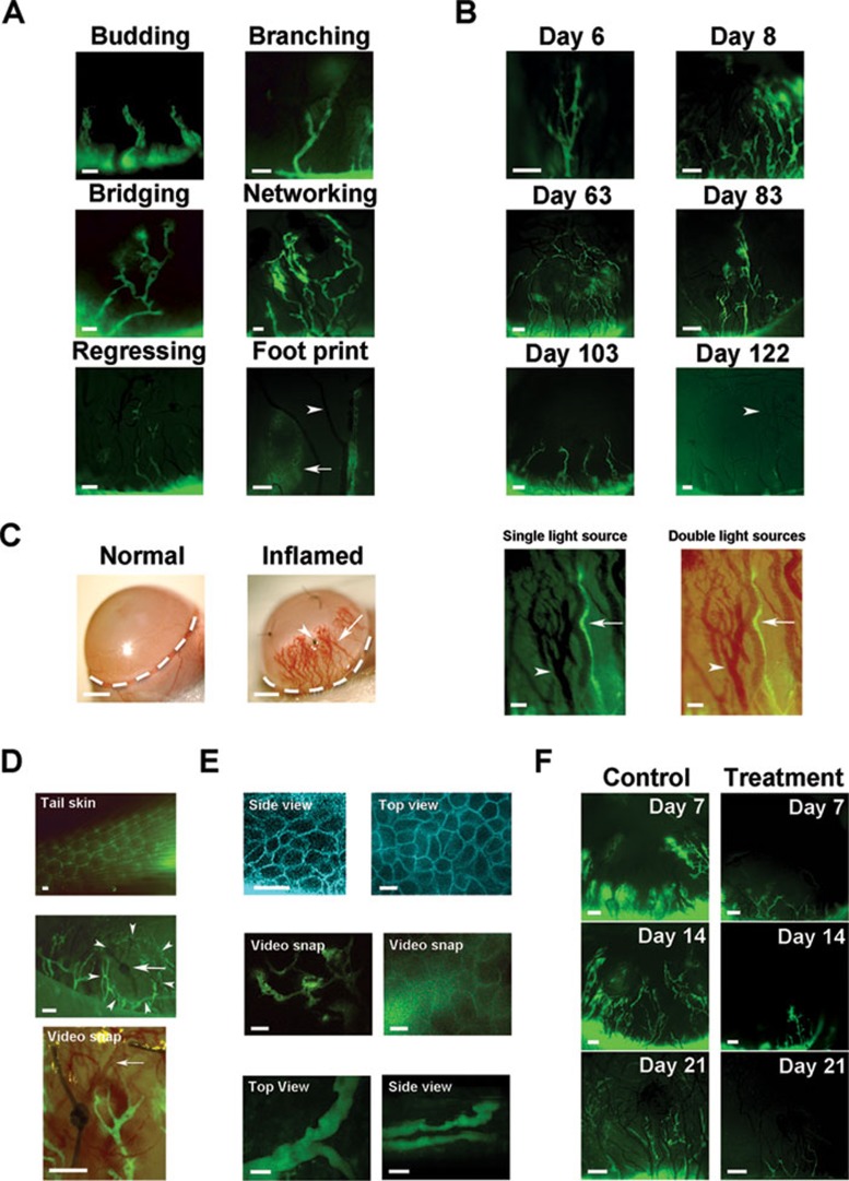
Figure 1.
Live imaging of newly formed lymphatic vessels in inflamed cornea. (A) Lymphatic vessels of various shapes and complexity. Arrow: footprints of an almost regressed lymphatic vessel; Arrowhead: blood vessel. (B) Longitudinal tracking of dynamic lymphatic processes in the same cornea in the progression (top panels) or regression phase (middle and lower panels). Arrowhead: blood vessel. (C) Comparison between ophthalmic slit-lamp microscopy (left two panels; scale bars: 1 000 μm) and the new imaging system, which also reveals lymphatic vessels (right two panels) in the corneas 2 weeks after suture placement. Right two panels: Arrows, lymphatic vessels; Arrowheads, blood vessels. (D) Comparison between live imaging of lymphatic vessels in normal tail skin and inflamed cornea 2 weeks after suture placement. Top: cross-sectional view of background lymphatics in the skin. Middle: entire morphological tree of new lymphatic vessels in the cornea showing terminal lymphatics (arrowheads) encircling a suture spot (arrow). Lower: snap picture (Supplementary information, Video S1) showing rapid red blood cell flow (arrow) inside new blood vessels. (E) Advanced two-photon live images showing ultra-structure of the cornea with lymphatic vessels. Top: side and top views of epithelial cells in layers. Middle: snap pictures showing lymphatic vessels in the stroma. Left (Supplementary information, Video S2): low magnification view taken along the central-to-peripheral axis. Right (Supplementary information, Video S3): high magnification view taken along the superficial-to-deep axis with lymphatic vessels behind the epithelium. Lower: snap pictures (Supplementary information, Video S4) showing two lymphatic vessels that appeared to cross over each other from the top view (left) were located at different layers from a side view (right). Pictures are assigned pseudo colors. Scale bars: 20 μm. (F) Real time in vivo evaluation of the effect of systemic VEGFR-2 blockade on corneal inflammatory lymphangiogenesis. Scale bars: 100 μm unless otherwise indicated.

