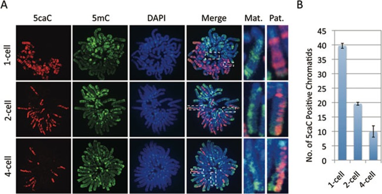
Figure 4.
Replication-dependent loss of 5caC during mouse preimplantation development. (A) Representative images of mitotic chromosome spreads co-stained with 5caC (red), 5mC (green) antibodies or DAPI (blue) at 1-cell, 2-cell, and 4-cell stages as indicated. Shown are the images of chromosomes from one blastomere at each developmental stage. Note that 5caC is present in both chromatids of the sperm-derived chromosomes at 1-cell stage embryos. However, only one of the two chromatids of the sperm-derived chromosomes stained positively for 5caC at the 2-cell stage, and only about a quarter of the sister chromatids stained positively for 5caC at the 4-cell stage. The dotted squares represent the enlarged paternal (Pat.) or maternal (Mat.) chromosomes, respectively. (B) Quantification of the total numbers of 5caC positive chromatids in each blastomere. The quantification method is the same as that described in Figure 3B. The experiments were repeated for four times and the total number of blastomeres examined at the 1-cell, 2-cell, and 4-cell stages were 19, 24, and 27, respectively. Bars represent standard deviation.

